
Figure 2. Flow velocity and transit blood flow volume in WT and Bmal1-KO mice.
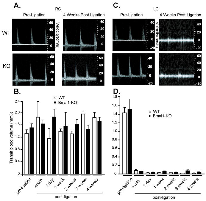
Doppler flow velocity in common carotid arteries was determined by echo ultrasound using PWD Mode imaging (see methods) in RC (A) and LC pre- and 4 weeks post-ligation (C). Transit blood flow was then determined in the RC (B) and LC (D) in weekly intervals to 4 weeks post-ligation. B. Contralateral, unligated RC did not display significant changes in WT versus Bmal1-KO mice before nor after the surgical ligation of LC (N=6/group). D. Blood flow in LC artery underwent a dramatic drop after ligation which was constant and not different in WT versus Bmal1-KO mice over the course of 4 weeks after ligation (N=6/group).