
Figure 8. Impaired response to batimastat in Bmal1-KO mice.
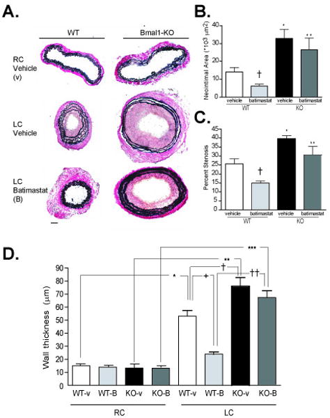
A. Von Gieson staining of elastin fibers revealed enhanced intimal hyperplasia in the left common carotid artery of Bmal1-KO mice undergoing LC ligation for 4 weeks. Neointima formation and wall thickening was partly prevented in WT mice administered batimastat. Neointimal area, wall thickness, and lumen diameter were calculated by morphometry of cross sections (see methods). Typical vascular remodeling evident as a lumen reduction was evident after 4 weeks of arterial ligation in wild-type mice, while Bmal1-KO mice exhibited no change in diameter. Analysis revealed an increase in neointimal area (B) and percent stenosis (C) in Bmal1-KO mice administered vehicle i.p. (N=3 *p<0.05). Neointima formation and stenosis were reduced in batimastat-treated WT mice compared to vehicle-treated WT ((N=3 *p<0.05, unpaired T test) while this effect was abrogated in Bmal1-KO mice. Wall thickness (D) (calculated at the level of EEL) was increased in both WT and Bmal1-KO ligated LC compared to RC control with the LC Bmal1-KO being significantly larger than the control LC WT. Batimastat treatment significantly reduced media thickness in LC WT compared to vehicle control LC WT, but again did not have any significant effect in LC of Bmal1 KO treated mice (N=3 *p<0.05, one-way ANOVA, Tukey post-test).