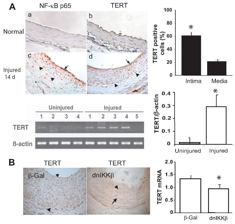
Figure 1. Activation of TERT expression in the injured artery is NF-κB dependent.
A: Top panel left: Immunohistochemistry for TERT and NF-κB p65 in the rat carotid artery. The arrowheads indicate internal elastic lamina, and the arrows indicate cells showing representative positive signals for TERT and p65, respectively (brown color). (Original magnification: X400). Lower panel left: RT-PCR analysis of TERT and β-actin from uninjured and injured carotid arteries. B: Left panel: Immunohistochemistry for TERT in artery transduced with adenovirus carrying either E coli β -Galactosidase (β-Gal) or dominant-negative mutant of IκB kinase β (dnIKKβ) at day 14, the arrow heads indicate TERT positive cells and the arrows indicate internal elastic lamina. Right panel: qRT-PCR analysis of TERT from the injured carotid arteries transduced with β-Gal or dnIKKβ at day 14, respectively (n=5 to 7). TERT transcripts normalized to Hprt, data are presented as mean ± SEM *p<0.05.

