Abstract
Selective peripheral nerve transfers represent an emerging reconstructive strategy in the management of both pediatric and adult brachial plexus and peripheral nerve injuries. Transfer of the lateral antebrachial cutaneous nerve of the forearm into the distal ulnar nerve is a useful means to restore sensibility to the ulnar side of the hand when indicated. This technique is particularly valuable in the management of global brachial plexus birth injuries in children for which its application has not been previously reported. Four children ages 4 to 9 years who sustained brachial plexus birth injury with persistent absent sensibility on the unlar aspect of the hand underwent transfer of the lateral antebrachial cutaneous nerve to the distal ulnar nerve. In three patients, a direct transfer with a distal end-to-side repair through a deep longitudinal neurotomy was performed. In a single patient, an interposition nerve graft was required. Restoration of sensibility was evaluated by the “wrinkle test.”
Keywords: Lateral antebrachial cutaneous nerve, Brachial plexus birth injuries, Wrinkle test, Restore sensibility
Introduction
Selective peripheral nerve transfers represent a great advancement in the treatment of brachial plexus and peripheral nerve injuries. Improved understanding in regional peripheral nerve anatomy and intraneural topography has confirmed redundancy in motor fascicles and innervation territories of sensory nerves [1]. As a result, several motor and sensory nerve transfers have been described [1–4].
Sensory loss on the ulnar border of the hand and in the small and ring fingers creates debilitating losses in protective hand sensation in patients with global plexus, lower trunk, or terminal branch injuries. Trophic ulcerations, burns, and self-mutilation may be seen. Reconstruction of ulnar sensory loss in the hand and wrist most commonly involved transfer of intact median nerve fascicles in the distal forearm to the ulnar sensory nerve and dorsal sensory branch of the ulnar nerve [2–8]. Specifically, median nerve sensory fibers in the distal forearm may be directly coapted to the ulnar sensory nerve at the distal forearm and the dorsal sensory branch of the ulnar nerve using side-to-end/terminolateral neurorrhaphies through adjacent epi/perineurial windows in the ulnar side of the median nerve [2–6]. Additionally, median nerve common sensory fascicles to the third web space may be coapted to the ulnar nerve common sensory branch to the fourth web space and the ulnar digital nerve to the small finger.
Oberlin et al. [9] recently described transfer of the proximal lateral antebrachial cutaneous nerve [LABCN] into the dorsal sensory branch of the ulnar nerve using a 15 cm sural nerve interpositional graft in two adult patients with lower plexus injuries. In these two patients, protective sensation was restored on the ulnar border of the hand. In contrast, we describe direct LABCN transfer into the ulnar nerve at the distal forearm using deep longitudinal neurotomy in order to restore protective sensation in the ulnar side of the hand in children with persistent ulnar sensory deficits despite prior plexus reconstructions for global injuries. Our experience with this sensory nerve transfer in four consecutive children operated on by the same senior surgeon (JAIG) presented.
Case 1 (TV)
A 5-year-old girl was seen for consideration of reconstructive surgery for multiple hand sequelae resulting from a global left brachial plexus birth injury. The total absence of sensibility of the hand was particularly debilitating. At age 6, she underwent a transfer of the lateral antebrachial cutaneous nerve into the ulnar nerve using a 6-cm interpositional graft of the superficial radial nerve. No protective sensibility had been recovered when seen in follow-up at 9 years of age.
Case 2 (JM)
A 4-year-old boy presented with persistent sequelae involving the hand as a result of a global right brachial plexus injury. As an infant, he had undergone a brachial plexus neurolysis at another institution and experienced limited recovery. Persistent lack of ulnar sensibility resulted in self-mutilation. At age 7, he underwent a direct transfer of the lateral antebrachial cutaneous nerve into the ulnar nerve in the distal forearm through a deep longitudinal neurotomy. Concomitant transfer of the flexor carpi ulnaris into the extensor carpi radialis brevis was performed to restore active wrist extension. At 6 months after surgery, he demonstrated protective sensibility on the ulnar aspect of the hand with a positive “wrinkle test” [10, 11] of the volar aspects of the ring and small fingertips that was not present on preoperative assessment.
Case 3 (CL)
A 6-month-old boy underwent exploration and microsurgical neurolysis with extensive nerve grafting for a global right brachial plexus injury. Postoperatively, persistent absence of sensibility on the ulnar side of the hand resulted in chronic self-mutilation of the small finger. At age 5, he underwent a lateral antebrachial cutaneous to ulnar nerve transfer. At follow-up 5 years after the nerve transfer, he had recovered protective sensibility on the ulnar aspect of the hand and a positive wrinkle test of the volar pad of the small finger was documented [10]. No further episodes of self-mutilation occurred.
Case 4 (EV)
A 7-month-old boy underwent reconstruction of a global right brachial plexus injury. Multiple pseudomeningoceles were noted on preoperative MRI. He had progressive recovery of shoulder and extremity function. At age 3.5 years, he underwent reconstruction of an internal rotation contracture of the shoulder with a subscapularis slide and muscle transfer. At age 4.5 years, he underwent a biceps rerouting for correction of a supination deformity of the forearm. By 5 years of age, it was observed that his hand function was compromised by repeated self-mutilation due to the absence of sensibility on the unlar side. At 6.5 years of age, he underwent transfer of the right lateral antebrachial cutaneous nerve to the right unlar nerve. The postoperative period was uneventful. One year after the surgery, he reported subjective sensibility in the small finger and had a positive wrinkle test. No further episodes of self-mutilation have been noted.
Surgical Technique
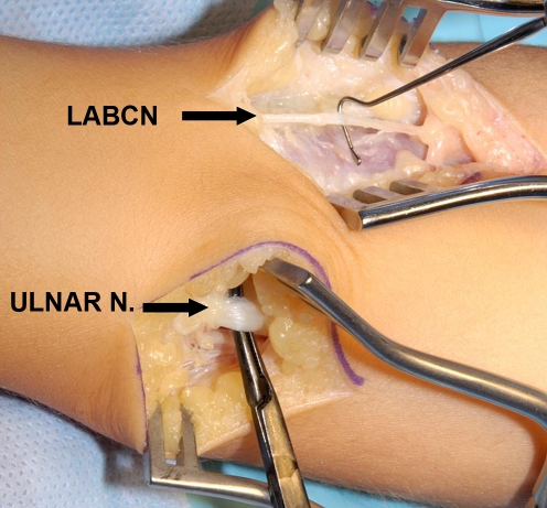
The lateral antebrachial cutaneous nerve of the forearm represents the terminal extension of the musculocutaneous nerve. It lies between the brachialis and biceps brachii muscles, and enters the proximal forearm lateral to the biceps tendon. A 5-cm linear incision over the palmar–radial aspect of the proximal forearm just distal to the elbow flexion skin crease is utilized. Following the superficial surgical dissection, the LABCN is identified just deep to the investing fascia of the forearm alongside the cephalic vein. Awareness of anatomic variations is necessary [12]. The LABCN is mobilized as distal as possible, and if proximal arborization is identified, the largest of the branches is selected and mobilized as distal as possible (Fig. 1). Smaller branches are divided using bipolar cautery. Loupe magnification and a lighted retractor aid in limiting the length of the necessary incision.
Figure 1.
Exposure of adequate length of LACN and ulnar nerve.
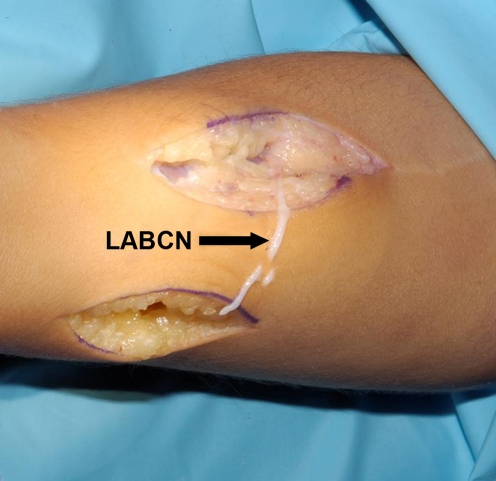
A separate longitudinal volar–ulnar incision is made in the distal forearm over the ulnar neurovascular bundle. When concomitant tendon transfers are performed (i.e., reconstruction of active wrist extension), a single incision may be considered. Following mobilization of the LABCN, a subcutaneous tunnel is developed between the proximal radial and distal ulnar exposures. The mobilized LABCN is divided as distal as possible and is passed through the tunnel to the main ulnar nerve trunk in a tension-free position. Utilizing standard microsurgical technique, a deep longitudinal neurotomy into the ulnar nerve is created proximal to the take-off of the dorsal ulnar sensory branch. The LABCN is placed into the neurotomy and the juncture completed with fibrin glue (Fig. 2).
Figure 2.
Divided LACN prior to passage through the subcutaneous tunnel and transfer.
Discussion
Although sensory nerve transfers are less widely used than motor nerve transfers, the principles of application are similar. The donor nerve should be in proximity to the recipient nerve’s sensory end organ receptors. The donor sensory nerve should innervate only a noncritical sensory territory, and the donor nerve should be a pure sensory nerve.
Mobilization of the LABCN as far distal as possible should allow for a tension-free terminolateral coaptation at the deep neurotomy site within the main ulnar nerve trunk at a site proximal to the take-off of the dorsal ulnar sensory branch. Familiarity with the anatomic course of the lateral antebrachial cutaneous nerve facilitates execution of this transfer. The LABCN has been shown to lie in close proximity to the cephalic vein in zone 1 (i.e., from the interepicondylar line of the elbow to the proximal crossover of the abductor pollicus longus and extensor pollicus brevis with the extrinsic radial wrist extensors) of the forearm within the subcutaneous adipose layer after piercing the investing fascia between the brachialis and biceps at the level of the elbow [12, 13]. We therefore advocate identification and neurolysis of the LABCN from proximal to distal, beginning at the level of antecubital fossa where it is more superficial and reliably distinguishable from the superficial radial nerve (SRN). Distally (i.e., zone 2 of the forearm), the SRN and LABCN arborize and run in the same tissue plane.
Anatomic studies [14–16] have confirmed the significant and constant anatomic overlap of the LABCN and SRN with regard to sensory territory innervation as a result of neural plexus interconnections between these two sensory nerves at the level of the wrist. In addition, the LABCN is most often only a minor contributor to the dorsal digital nerves of the thumb [14]. These anatomic data support the use of the LABCN as a select sensory nerve donor as minimal sensory disturbance is expected following its use.
An additional advantage of the direct transfer of the LABCN into the ulnar nerve sensory nerve in the distal forearm (and the alternative technique described by Oberlin and colleagues [9]) is that this transfer avoids use of the median nerve as a sensory donor and thus eliminates the potential for creating an additional sensory disturbance in the hand. While an end-to-end neurorrhaphy is preferred with motor nerve transfers, end-to-side (i.e., terminolateral) neurorrhaphies are acceptable with sensory nerve transfers.
Our technique is to create a deep longitudinal neurotomy using a diamond knife under high magnification (×10). Both perineureium and epineurium are incised, and some fibers undoubtedly opened as well.
Because this transfer is reported in young children, recovery of sensibility is much more rapid than would be expected in adults. Longer-term follow-up is needed to determine if the protective sensibility recovered topographically matches the recipient nerve zone [17]. As with other sensory nerve transfers, it is possible that the sensory feedback is perceived in the topography of the donor nerve. Reinnervation of the volar pulp is, however, confirmed by the positive wrinkle test.
In summary, the three children who underwent direct LABCN to ulnar nerve transfer utilizing a deep neurotomy in the distal forearm recovered protective sensation to the ulnar side of the hand and demonstrated objective evidence of ulnar nerve territory sensory reinnervation as signaled by the positive wrinkle test [10] in the pad of the small finger at latest follow-up. The poor result was likely multifactorial. Extensive forearm scarring and perineural fibrosis limited mobilization of the LABCN, and as a result, a long interpositional graft was required. Perhaps the quality of the superficial radial graft also compromised the result. Direct transfer of the LABCN offers reliable protective reinnervation in the sensory territory of the ulnar nerve in the hand without sacrificing a critical area of extremity sensibility.
Acknowledgments
Conflict of interest disclosure The authors declare that they have no conflict of interest.
Footnotes
Investigation performed at the NYU Hospital for Joint Diseases, New York, NY, and Miami Children’s Hospital, Miami, FL.
References
- 1.Tung TH, Mackinnon SE. Nerve transfers: indications, techniques, and outcomes. J Hand Surg Am. 2010;35:332–341. doi: 10.1016/j.jhsa.2009.12.002. [DOI] [PubMed] [Google Scholar]
- 2.Nath RK, Mackinnon SE. Nerve transfers in the upper extremity. Hand Clin. 2000;16(1):131–139. [PubMed] [Google Scholar]
- 3.Mackinnon SE, Novak CB. Nerve transfers. New options for reconstruction following nerve injury. Hand Clin. 1999;15(4):643–666. [PubMed] [Google Scholar]
- 4.Mackinnon SE, Colbert SH. Nerve transfers in the hand and upper extremity surgery. Tech Hand Up Extrem Surg. 2008;12(1):20–33. doi: 10.1097/BTH.0b013e31812714f3. [DOI] [PubMed] [Google Scholar]
- 5.Brown JM, Mackinnon SE. Nerve transfers in the forearm and hand. Hand Clin. 2008;24(4):319–340. doi: 10.1016/j.hcl.2008.08.002. [DOI] [PubMed] [Google Scholar]
- 6.Battiston B, Lanzetta M. Reconstruction of high ulnar nerve lesions by distal double median to ulnar nerve transfer. J Hand Surg Am. 1999;24(6):1185–1191. doi: 10.1053/jhsu.1999.1185. [DOI] [PubMed] [Google Scholar]
- 7.Ihara K, Doi K, Sakai K, Kuwata N, Kawai S. Restoration of sensibility in the hand after complete brachial plexus injury. J Hand Surg Am. 1996;21(3):381–386. doi: 10.1016/S0363-5023(96)80348-0. [DOI] [PubMed] [Google Scholar]
- 8.Matloubi R. Transfer of sensory branches of radial nerve in hand surgery. J Hand Surg Br. 1988;13(1):92–95. doi: 10.1016/0266-7681(88)90062-9. [DOI] [PubMed] [Google Scholar]
- 9.Oberlin C, Teboul F, Severin S, Beaulieu JY. Transfer of the lateral cutaneous nerve of the forearm to the dorsal branch of the ulnar nerve, for providing sensation on the ulnar aspect of the hand. Plast Reconstr Surg. 2003;112(5):1498–1500. doi: 10.1097/01.PRS.0000080583.35200.53. [DOI] [PubMed] [Google Scholar]
- 10.O’Riain S. New and simple test of nerve function in hand. Br Med J. 1973;3(5881):615–616. doi: 10.1136/bmj.3.5881.615. [DOI] [PMC free article] [PubMed] [Google Scholar]
- 11.Smith P. Lister’s the hand: diagnosis and indications. 4. London: Churchill Livingstone; 2002. pp. 505–512. [Google Scholar]
- 12.Beldner S, Zlotolow DA, Melone CP, Jr, Agnes AM, Jones MH. Anatomy of the lateral antebrachial cutaneous and superficial radial nerves in the forearm: a cadaveric and clinical study. J Hand Surg Am. 2005;30(6):1226–1230. doi: 10.1016/j.jhsa.2005.07.004. [DOI] [PubMed] [Google Scholar]
- 13.McFarlane RM, Mayer JR. Digital nerve grafts with the lateral antebrachial cutaneous nerve. J Hand Surg Am. 1976;1(3):169–173. doi: 10.1016/s0363-5023(76)80034-2. [DOI] [PubMed] [Google Scholar]
- 14.Mok D, Nikolis A, Harris PG. The cutaneous innervation of the dorsal hand: detailed anatomy with clinical implications. J Hand Surg Am. 2006;31(4):565–574. doi: 10.1016/j.jhsa.2005.12.021. [DOI] [PubMed] [Google Scholar]
- 15.Mackinnon SE, Dellon AL. The overlap pattern of the LABCN and SRN. The overlap pattern of the lateral antebrachial cutaneous nerve and the superficial branch of the radial nerve. J Hand Surg Am. 1985;10(4):522–526. doi: 10.1016/s0363-5023(85)80076-9. [DOI] [PubMed] [Google Scholar]
- 16.Abrams RA, Brown RA, Botte MJ. The superficial branch of the radial nerve: an anatomic study with surgical implications. J Hand Surg Am. 1992;17(6):1037–1041. doi: 10.1016/S0363-5023(09)91056-5. [DOI] [PubMed] [Google Scholar]
- 17.Stice RC, Wood MB. Neurovascular island skin flaps in the hand: functional and sensibility evaluations. Microsurgery. 1987;8:162–167. doi: 10.1002/micr.1920080311. [DOI] [PubMed] [Google Scholar]


