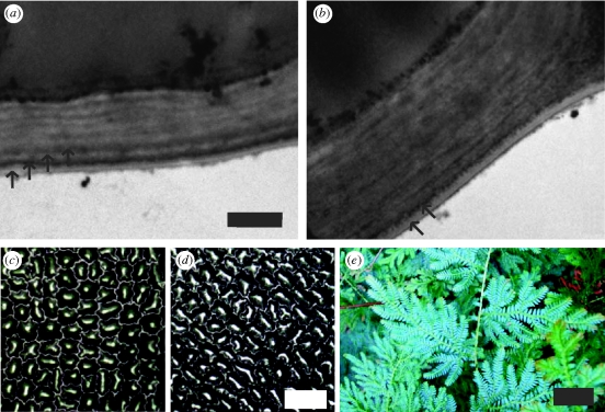
Figure 3.
TEM micrographs of the outer cell wall and the cuticle from the upper epidermis of (a) a juvenile blue leaf and (b) an older green leaf (scale bar, 500 nm). Arrows indicate the layers observed in the cuticle. Optical micrographs of surface morphology of plant cells for (c) a juvenile blue leaf and (d) a mature green leaf (scale bar, 50 µm). (e) Photograph of juvenile S. willdenowii leaves (scale bar, 30 mm).

