FIGURE 6.
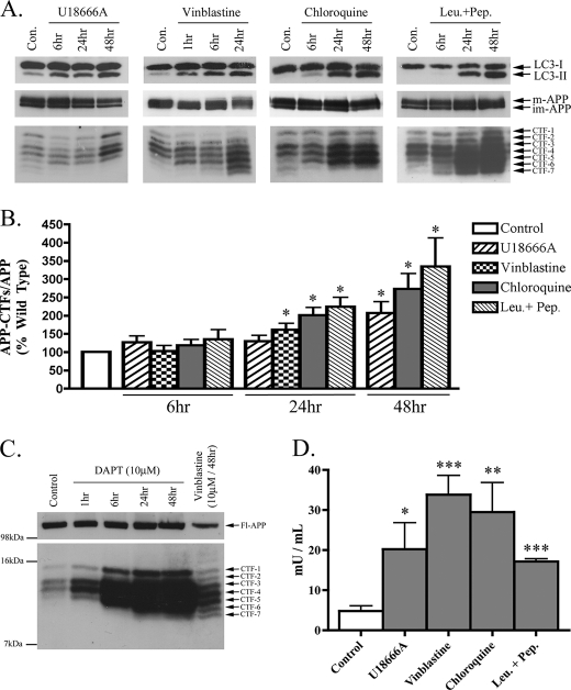
Impaired lysosomal flux causes AV and APP-CTF accumulation in primary cortical neurons. Rat primary cortical neurons (DIV14) were treated with U18666A (2 μg/ml), vinblastine (10 μm), chloroquine (10 μm), and leupeptin + pepstatin (Leu.+Pep.) (each at 20 μm) for the times shown. A, representative immunoblot images of LC3-I/II, APP, and APP-CTF expressions for each treatment condition are shown. B, histogram of APP-CTF/Fl-APP expression in primary neurons treated as in A, n = 6 for all conditions, mean ± S.E.). C, immunoblot confirming the specificity of the APP-CTFs detected in primary cortical neurons using the γ-secretase inhibitor, DAPT (10 μm). Importantly, the CTFs detected in vinblastine (10 μm, 48 h)-treated neurons perfectly co-migrated with those detected in cells treated with DAPT. D, histogram representing amounts of sequestered LDH activity in neurons treated for 24 h, n = 4, mean ± S.E.

