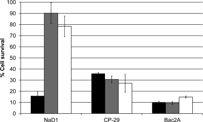
FIGURE 7.
Killing of hyphal cells after cell wall treatments. The death of untreated fungal hyphae (black bars) was compared with that of hyphae that had been treated with either proteinase K (gray bars) or β-glucanase (white bars). Both proteinase K and β-glucanase treatments prevented NaD1-induced cell death. In contrast, CP-29 and Bac2A killed treated hyphae as efficiently as the untreated control. All peptides were used at 5 μm, and error bars represent S.E. (n = 4).

