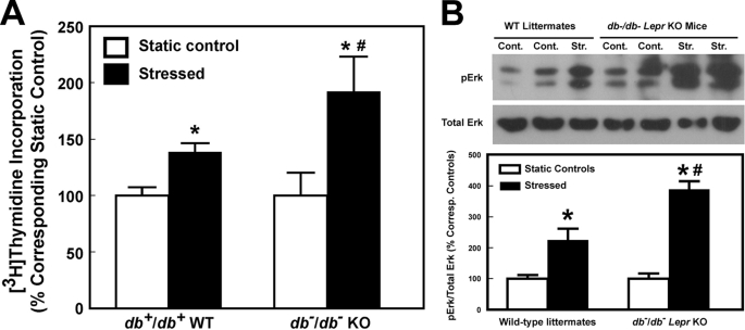
FIGURE 5.
Effects of fluid shear on [3H]thymidine incorporation (A) and Erk1/2 phosphorylation (B) of osteoblasts of 12-week-old female Lepr-deficient db−/db− mice and of osteoblasts of age- and sex-matched littermates. In A, results are shown as the percentage of respective static cells (mean ± S.D., n = 6). *, p < 0.05 versus static control; #, p < 0.05 versus the WT littermates. In B, the top shows a representative blot of pErk1 and pErk2 (using an anti-pErk1/2 antibody) and total Erk1/2 (identified by an anti-pan-Erk antibody). The bottom summarizes the relative amounts of pErk1/2 normalized against total Erk, and results are shown as a percentage of respective static control (mean ± S.D. (error bars), n = 4–6). *, p < 0.05, compared with static control; #, p < 0.05, compared with osteoblasts of WT littermates.

