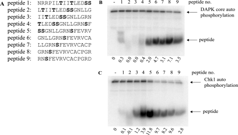
FIGURE 3.
The multiprotein docking peptide from the ubiquitin signal in the p53 DNA-binding domain contains an in vitro phosphoacceptor site. A, overlapping peptides corresponding to the conformationally flexible ubiquitination signal (with Ser260/261 phosphoacceptor sites in peptides 1–5 or Ser269 phosphoacceptor site in peptides 5–9) were incubated in kinase reactions containing [γ-32P]ATP and either DAPK core (B) or Chk1 (C) for 30 min at 30 °C. Reaction products were resolved via 20% SDS-PAGE and dried, and phosphopeptides were visualized by a PhosphorImager (highlighted by arrows). The intensity of peptide phosphorylation was quantified and normalized to kinase autophosphorylation and is expressed below each panel.

