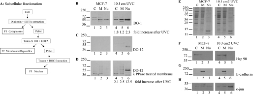
FIGURE 7.
Serine 269-phosphorylated p53 has a selective subcellular localization. A, schematic representation of the fractionation methodology. B–D, effects of irradiation on p53 phosphorylation at the DO-12 epitope in MCF7 cells. MCF-7 cells were untreated or irradiated with UVC and grown for a further 6 h before fractionation into cytosolic (C), membrane/organelle (M), and nuclear fractions (Nu). Fractions were resolved by electrophoresis and immunoblotting with DO-1 (B) and DO-12 before (C) and after phosphatase treatment of the membrane (D) to define p53 localization. Band intensity was quantified by Scion Image software, and the -fold increase in antibody signal following UVC irradiation is indicated below B and D, respectively. Equivalent sample loading is shown by Coomassie staining of the various fractions (E), and fractionation of the distinct subcellular fractions was demonstrated by probing for known cytoplasmic (Hsp90), membrane (E-cadherin), and nuclear (c-Jun) marker proteins (F, G, and H, respectively).

