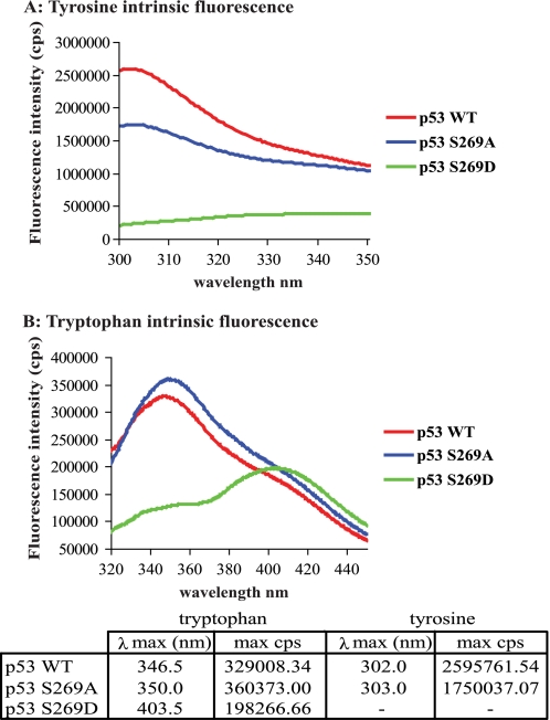
FIGURE 5.
Alanine or aspartate substitution of serine 269 generates conformational differences within the p53 core domain. Tyrosine (A) and tryptophan (B) fluorescence spectra of the wild type p53, p53S269A, and p53S269D core domain variants were determined and corrected for the presence of buffer background spectra as described previously (32). The fluorescent maxima for both tryptophan and tyrosine emission and the corresponding wavelength are detailed in the table below. cps, counts per second.

