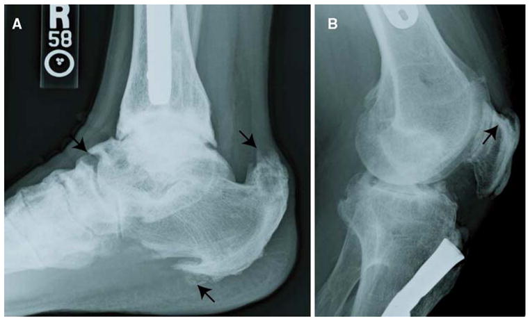
Fig. 1.

Enthesophyte formation in XLH patients. a Enthesophytes at the site of enthesis attachment to the plantar aspect of the calcaneus are evident. Prominent enthesopathy and progressive mineralization at the site of the Achilles tendon are marked by arrows. b Calcification of the patellar ligament insertion is shown by the arrow. Note the fusion of the patella to the femur
