
Figure 2.
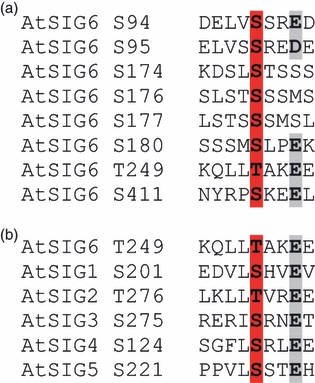
Alignment of analysed cpCK2 substrate motifs.The phosphoacceptor Ser or Thr residues are marked in red, and the acidic residues at n + 3 are marked in grey.(a) Multiple alignment of the eight cpCK2 substrate sites in AtSIG6 shown in Figure 1.(b) Alignment of the ‘general’ substrate site T249 from AtSIG6 with motifs from the conserved region of the other Arabidopsis sigma factors, AtSIG1– AtSIG5.