
Figure 3.
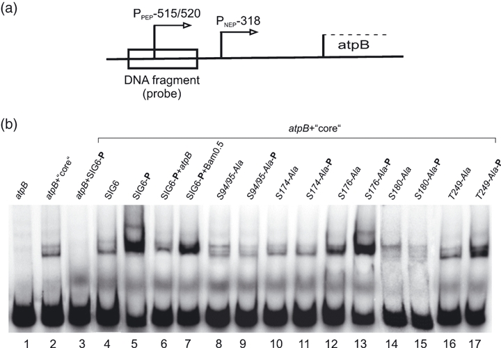
Phosphorylation-dependent DNA binding of recombinant AtSIG6 in vitro.(a) Map position of the DNA fragment used as a probe. The 424-bp fragment carries the atpB PEP-515/-520 promoter (Pfalz et al., 2006) and a 154-bp section downstream of the transcription start site.(b) EMSA experiments using promoter fragment, Escherichia coli core polymerase and recombinant sigma proteins, with or without prior phosphorylation by recombinant cpCK2. The free radiolabelled DNA fragment migrates at the bottom (lane 1; atpB). Core polymerase alone (lane 2) but not AtSIG6 alone (lane 3) is able to bind to the promoter fragment. Complete reactions containing probe, core enzyme and either phosphorylated or mock-phosphorylated sigma proteins are shown in lanes 4–17 (atpB + core). Lanes 6 and 7 show competition with a twofold molar excess of unlabelled promoter fragment (atpB), but not with a promoter-less fragment Bam0.5 at 10-fold excess. Like AtSIG6 itself, none of the mutagenized derivatives show sigma activity in the unphosporylated form (lanes 8, 10, 12, 14 and 16).