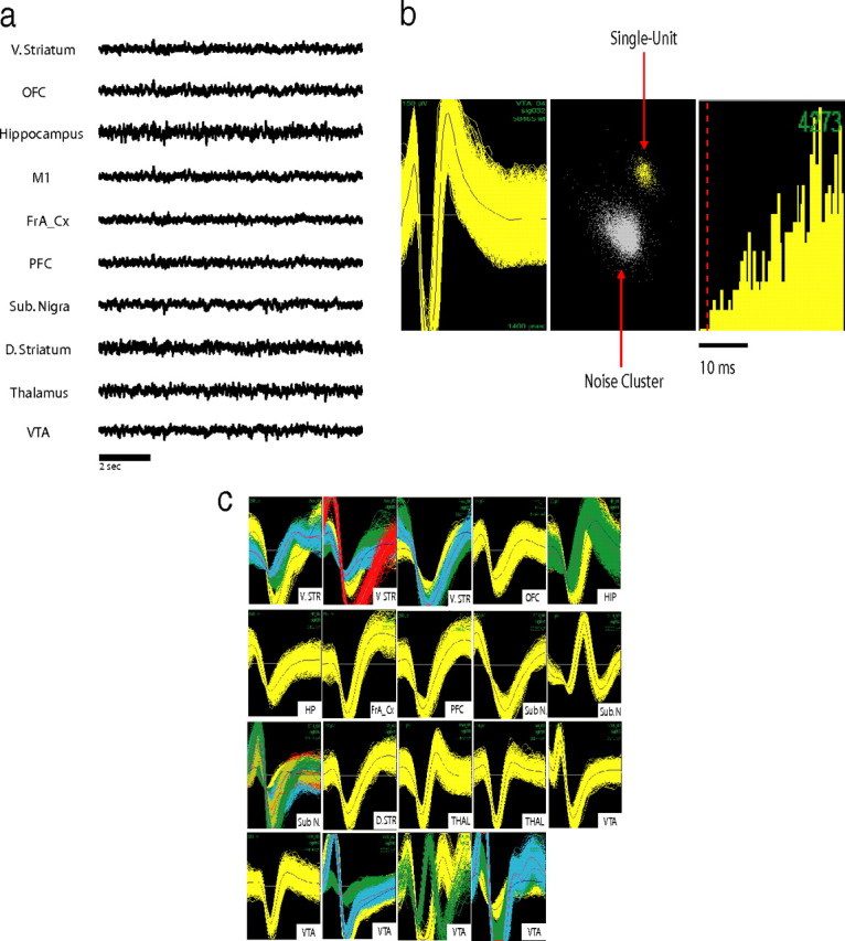
Figure 2.

Multisite electrophysiological recording in mice. a, LFP recordings from WT baseline waking period. LFPs were simultaneously recorded from ventral striatum (V. STR), frontal association cortex (FrA_Cx), hippocampus (HP), M1, OFC, PFC, substantial nigra (Sub. N), dorsal lateral striatum (D. STR), medial dorsal thalamus (THAL), and VTA. b, Example of an isolated single unit in VTA. From left to right, Depiction of the extracellularly recorded wave form of the unit (x-axis, 1400 ms; y-axis, 150 mV), interspike interval histogram, and projection of the clusters correspondent to the unit and the noise based on analysis of the first two principal components of the waveforms recorded (x-axis, PC1; y-axis, PC2). c, Simultaneous recording of 37 single units across nigrostriatal and mesolimbic brain circuits in an awake behaving mouse.
