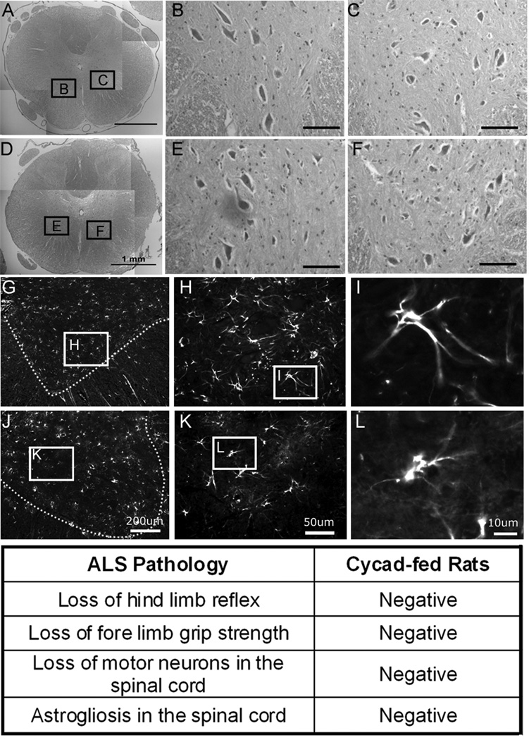
Figure 5.
Cycad-fed rats do not display amyotrophic lateral sclerosis (ALS) phenotype or pathology. Cohort 1A and 1B (flour, n = 7; cycad, n = 8) were used to verify a lack of ALS pathology in cycad-fed rats. Representative images of motor neurons from the ventral horn of the lumbar spinal cord stained with hematoxylin and eosin from cycad-fed rat IJ (D–F) and flour-fed rat QR (A–C). Results indicate no differences in size of intact motor neurons on either side. There was also no astrogliosis in the spinal cord of cycad-fed rats (G–I) as compared to flour-fed rats. Scale bars: A, D = 1mm; G, J = 200µm; B, C, E, F = 100µm; H, K = 50µm; I, L = 10µm.

