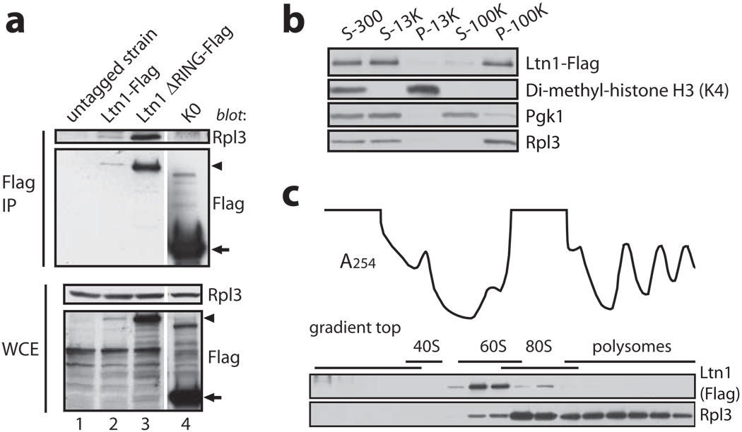
Figure 4. Ltn1 is predominantly associated with ribosomes.
Strains expressing C-terminally Flag-tagged endogenous Ltn1 (a-c) or Ltn1 ΔRING (a) were used in this figure. a, Ltn1 specifically co-IP’s with Rpl3. Indicated lysates were Flag IP’ed, followed by anti-Rpl3 blot. K0 and the untagged WT strain were negative controls. Arrowhead, Ltn1 and Ltn1 ΔRING; arrow, K0. b, Ltn1 is predominantly cytoplasmic. Pellet (P) and supernatant (S) samples were taken following centrifugation of lysate at 300, 13K and 100K × g. Blots were probed for Flag, Lys4-di-methylated histone H3, Pgk1, and Rpl3. c, Ltn1 is predominantly 60S-bound in steady-state. Ltn1’s distribution in sucrose gradient fractions analyzed by immunoblot. Line tracing, A254 profile.

