
Figure 6. Calcium activates mouse β-cell SK currents which significantly regulate β-cell membrane potential.
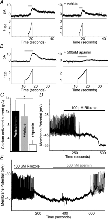
A, C57BL/6 mouse β-cell current recorded at −60 mV (top trace) and calcium change (lower trace) recorded during photolysis of caged calcium with the indicated pulse of 360 nm light (grey line); the right set of traces is from the same cell 5 min following vehicle treatment. B, C57BL/6 mouse β-cell current recorded at −60 mV (top trace) and calcium change (lower trace) recorded during photolysis of caged calcium with the indicated pulse of 360 nm light (grey line); the right set of traces is from the same cell 5 min following 100 nm apamin treatment. C, analysis of SK currents in response to vehicle or apamin (500 nm) (n = 4, +s.d., *P < 0.05). D, representative C57BL/6 islet electrical activity, induced with tolbutamide, in response to 100 μm riluzole (black bar). E, representative C57BL/6 islet electrical activity, induced with tolbutamide, in response to 100 μm riluzole (black bar) and 500 nm apamin (grey bar).