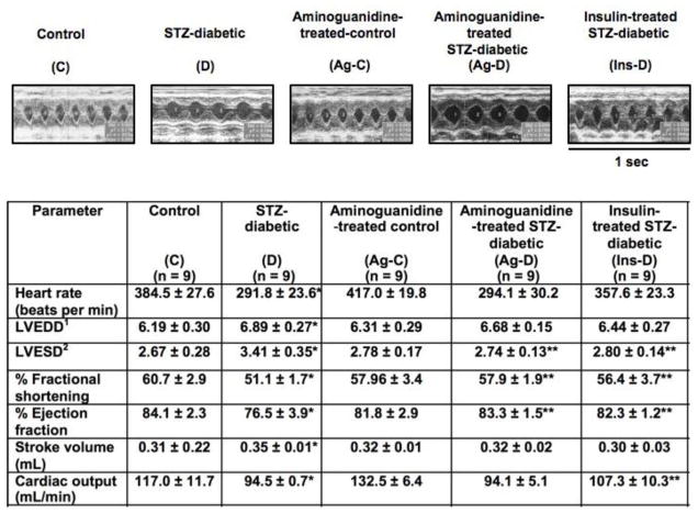
Figure 2.
Upper panels show representative M-mode echocardiograms from control (C), STZ-diabetic (D), Ag-treated control (Ag-C), Ag-treated STZ-diabetic (Ag-D) and insulin-treated STZ-diabetic (Ins-D) rats. Three loops of M-mode were captured for each animal. Values in table below are mean ± SEM (n ≥ 8). 1LVEDD - left ventricular end diastolic diameter. 2LVESD- Left ventricular end systolic diameter. * - Significantly different from controls (p<0.05). ** -Significantly different from STZ-diabetic (p<0.05).

