
Figure 7. JNK is activated in human liver fibrosis and promotes TGFβ and PDGF signaling in human hepatic stellate cells.
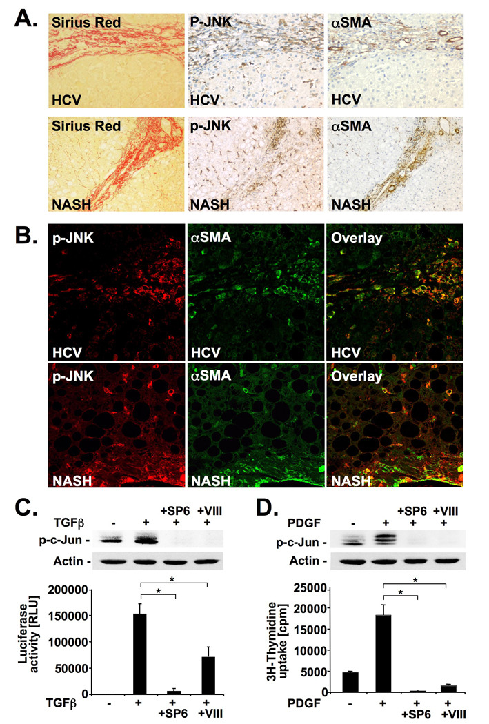
A–B. Serial sections of fibrotic livers from patients with HCV (n=3) or NASH (n=3) were stained for p-JNK, αSMA or by picrosirius red. Single sections were stained for αSMA (green) and p-JNK (red) followed by confocal microscopy. Shown are representative images from one patient each (B). C. Primary human HSCs were pretreated with SP600125 or JNK inhibitor VIII. c-Jun phosphorylation was determined by immunoblot 60 minutes after TGFβ (2 ng/ml) treatment (upper panel). (CAGA)9-driven luciferase was determined 6 hours after TGFβ (10 pg/ml) (lower panel). D. Primary human HSCs were pretreated with JNK inhibitors followed by PDGF (20 ng/ml) treatment. Phosphorylation of c-Jun (upper panel) and [3H]-thymidine incorporation (lower panel) were determined 15 minutes and 26h after PDGF stimulation, respectively. * p<0.05