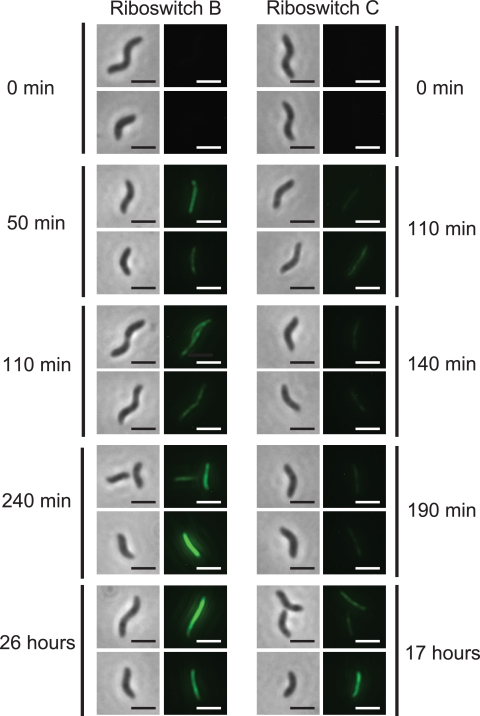
FIG. 2.
Images of riboswitches B and C controlling the expression of a MamK-GFP fusion in M. magneticum in the presence of theophylline (1 mM) as a function of time. Left panels are phase-contrast images; right panels monitor GFP fluorescence emission (2-μm scale bar). All fluorescent images were exposed for 6 s, and the cells were visualized with a 100× objective.

