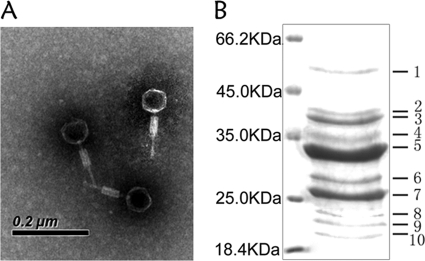
FIG. 1.
Characterizations of deep-sea thermophilic bacteriophage D6E. (A) Electron micrograph of D6E virions. Scale bar, 200 nm. (B) SDS-PAGE of proteins from purified D6E virions, followed by staining with Coomassie brilliant blue R250. The numbers indicate the excised bands for mass spectrometric analysis. The left lane contains the protein molecular mass marker (kDa).

