
Figure 5.
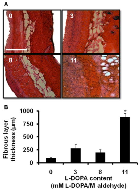
(A) Tissue fibrous layer thickness local to PEG:dextran variants increases with L-DOPA content. Mice were implanted with material (200 μL) subcutaneously and tissues were harvested after 9 days. Hematoxylin and eosin stained sections of tissue demonstrate increased cellular infiltration and formation of fibrous tissue surrounding the implanted material. Scale bar = 500 μm and applies to all images. (B) Fibrous layer thickness was measured and found to be significantly increased in 11 mM L-DOPA/M aldehyde conjugate. Error bars represent 1 standard error of measurement (n = 4 to 5). * indicates p < 0.05 determined by ANOVA with post hoc analysis when compared to unmodified PEG:dextran