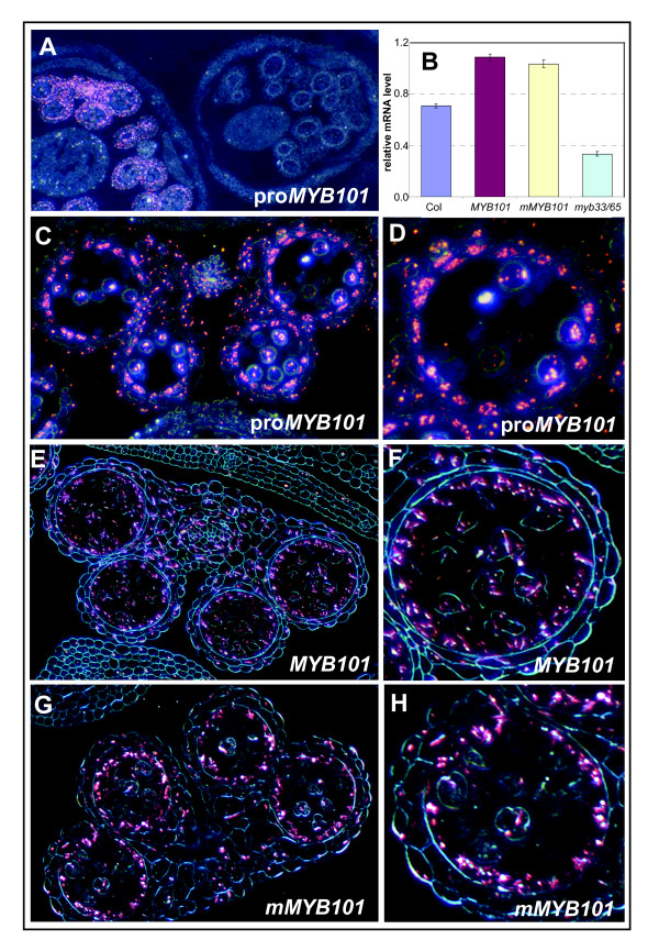
Figure 8.
MYB101 and mMYB101 transgenes have indistinguishable expression patterns. Anthers were stained overnight and embedded in paraffin. Transverse sections were examined by dark field microscopy. β-Glucuronidase (GUS) staining is shown by pink crystals. (A) Low magnification of proMYB101:GUS inflorescence showing MYB101 transcription is restricted to postmeiotic anthers. (B) Relative expression of MYB101 in wild type, MYB101/mMYB101 GUS lines and myb33.myb65. Analysis was performed on RNA extracted from inflorescences with measurements being the average of three replicates with error bars representing the standard error of the mean. mRNA levels are relative to cyclophilin. (C) GUS staining in proMYB101:GUS anthers. (D) Detail of a single locule of proMYB101:GUS. (E) GUS staining in MYB101:GUS anthers. (F) Detail of a single locule of MYB101:GUS. (G) GUS staining in mMYB101:GUS anthers. (H) Detail of a single locule of mMYB101:GUS.

