
Figure 2. Antigen presentation in the liver results in T cell tolerance.
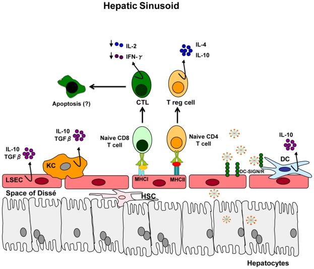
The liver sinusoid is lined by a fenestrated endothelium (liver sinusoidal endothelial cells, LSEC). Kupffer cells (KCs) and immature dendritic cells (DCs) are found in the sinusoids. Hepatic stellate cells (HSC) are located in the sub-endothelial space, known as the Space of Dissé. T cells that recognize antigen in the liver are exposed to immunosuppressive cytokines (IL-10 and TGF-β) that are synthesized by KCs, LSECs and DCs. Interaction of naïve T cells with LSEC results in differentiation of T cells into CD4+ regulatory T cells and impaired cytotoxic CD8+ T cells, followed by cell death. Hepatotropic viruses appear to be captured by DCs and/or LSECs in process that probably involves DC-SIGN or DC-SIGNR (for HCV) or other not yet defined cell-surface molecules (for HBV) for subsequent transfer to the underlying hepatocytes or viral particles may be internalized by hepatic DCs and LSECs for processing and presentation to naïve T cells (Adapted from 116 and 133).