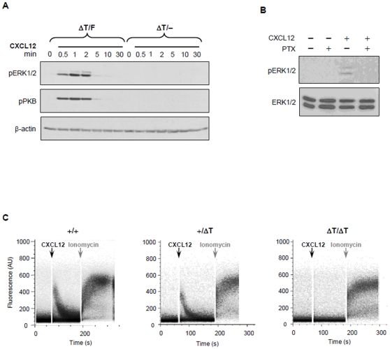
Figure 6. Impaired signaling pathways in ΔT cells.
(A) Splenocytes from conditional ΔT mice and their WT littermates were stimulated with 0.5 µg/ml CXCL12 for indicated times and immunoblot analysis for phosphorylated PKB at serine 473 (pPKB) or phosphorylated ERK (pERK) were performed. Equal loading of protein was confirmed by anti-β-actin staining. Results are representative of at least 3 independent experiments. (B) WT splenocytes, pretreated with 100 ng/ml of PTX for 1 h, were stimulated with 0.5 µg/ml of CXCL12 for 1 min and lysates immunoblotted for pERK and re-probed with antibody against total ERK1/2 for loading control. (C) FL cells from WT, heterozygous and homozygous ΔT embryos were collected and loaded with Fluo-4 and Fura-red calcium indicator dyes. Cells were stimulated with 0.5 µg/ml CXCL12 for 2 min followed by 2 µM ionomycin. Results are representative of 3 independent experiments.

