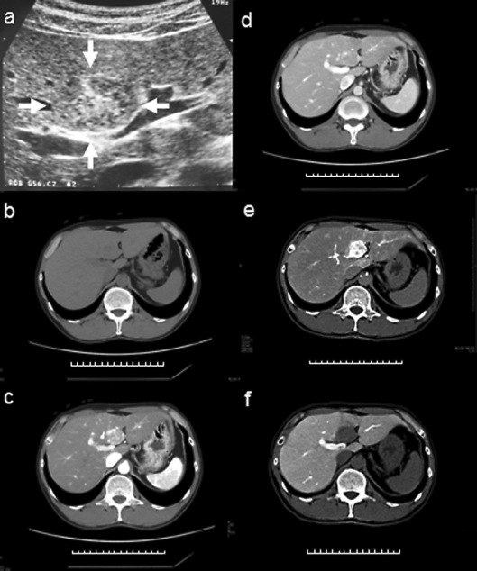
Fig. 1.
a Abdominal US revealed a well-circumscribed, heterogeneous hypoechoic tumor (arrow) with a central hyperechoic area. The tumor measured 34 × 24 mm and was located in segment 4 of the liver. b A precontrast CT scan showed that the lesion was a homogenous well-defined nodule with slightly low attenuation. The tumor had a diameter of 27 mm, and no fatty attenuation was visible within the lesion. c, d Contrast-enhanced CT revealed a hypervascular tumor in the arterial phase (c) and moderate washing out of the contrast medium in the portal phase (d). e, f A hypervascular tumor was observed on CT hepatic arteriography (e) and complete washing out of the contrast medium was observed on CT arterioportography (f).

