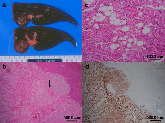
Fig. 3.
a The cut surface of the resected tumor appeared yellowish to tan and round with clear margins. No capsule was seen, and the diameter of the tumor was 28 mm. b, c Hematoxylin and eosin staining of the tumor showed that the tumor was almost exclusively composed of epithelioid smooth muscle cells that exhibited a trabecular growth pattern, few thick-walled blood vessels (arrow), and few adipose cells (b). The fat component accounted for only 5% of the largest cut surface area of the tumor (c). d On immunohistochemical analysis, the epithelioid smooth muscle cells were positive for HMB-45.

