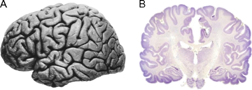
Fig. 3.
Two views of the human brain. a. Lateral view (rostral end is left, caudal is right) shows an apparently uniform surface marked by gyri and sulcal folds (Right hemisphere of J. Piłsudski’s brain, lateral view, image in the public domain). b. Coronal cross-section (cut at approximately the level of the dotted line in A) stained for cell bodies that mark neurons. The neocortex is the thin mantel layer (dark purple) on the surface of the brain. The white areas are connecting fiber pathways. Image reproduced with permission from http://www.brains.rad.msu.edu which is supported by the U.S. National Science Foundation. Images obtained with permission from Wiki Commons, http://commons.wikimedia.org/wiki

