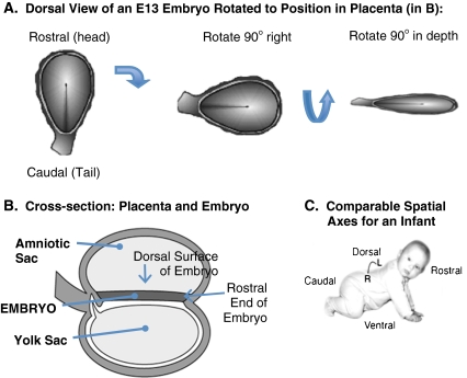
Fig. 4.
The major spatial dimensions of the E13 embryo. a. The dorsal surface view of the embryo on E13 is shown in the first panel. The wall of the amniotic sac has been cut away to reveal the dorsal surface (epiblast layer) of the embryo. The rostral (“head”) end of the embryo is on the top of this figure, and the caudal (“tail”) end is at the bottom. b. A lateral cross-section of the embryo and placenta at E13. On E13, the two-layered embryo is located centrally between two major placental sacs. The amniotic sac (which later in development will surround the embryo) is located above the embryo, and the yolk sac is located below. The rostral end of the embryo is to the right in this figure. To place the embryo shown in the first panel of A within the context of the lateral view of the embryo and placenta shown in B, it is necessary to first rotate the embryo so that the rostral end faces right (second panel of A), and then rotate the embryo in depth so that the dorsal surface faces up (last panel of A). C. The comparable rostral-caudal and dorsal-ventral spatial axes of an infant. The spatial axes of a crawling infant are comparable to the position of the embryo in B. Illustrations by Matthew Stiles Davis reprinted by permission of the publisher from THE FUNDAMENTALS OF BRAIN DEVELOPMENT: INTEGRATING NATURE AND NURTURE by Joan Stiles, Cambridge, Mass.: Harvard University Press, Copyright © 2008 by the President and Fellows of Harvard College

