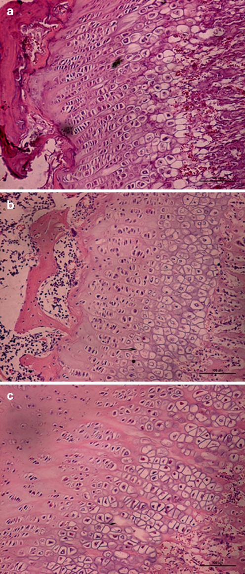
Fig. 1.
Epiphyseal plate showing cell necrosis (arrows) stained by haematoxylin and eosin (magnification ×200). a Normal control group (fourth week) rat epiphyseal cartilage cells, cell columns were arranged regularly, nucleus clear, transparent cytoplasm. b Normal feed + T-2 toxin group (fourth week), cell columns with disorders, mast cell layer condensation nuclei can be seen with pyknosis and lysis of nuclei, cells appear with vacuolated changes. c Low nutrition feed + T-2 toxin group (fourth week), cell columns with disorders, sparse, patchy cell-free zone appears. Lamellar necrosis in hypertrophic zone

