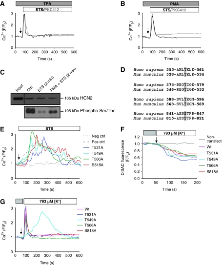
Figure 5.
Dephosphorylation of Thr549 is essential for the prolonged Ca2+ influx through HCN2. U1810 cells were pretreated with the PKC activators (A) 1 μM TPA or (B) 300 nM PMA, before exposure to STS or PKC412. (C) The HCN2 channel was immunoprecipitated and analysed by western blot for phosphorylation on Ser/Thr residues. (D) Conserved putative PKC phosphorylation sites in the C terminal of human and mouse HCN2 are marked in grey. (E) The mutant and wild-type forms of the HCN2 channel were expressed in HEK293 cells, which were subsequently loaded with Fluo-4/AM, and the intracellular Ca2+ level was monitored upon exposure to STS. Untransfected HEK293 cells served as negative control and cells transfected with wild-type HCN2 channel was used as a positive control. MitoDsRed was used as a transfection marker. Hyperpolarization was induced by decreasing the K+ level from 4.7 mM (grey box) to 783 μM (white box), and polarization was monitored using DiBAC (F) and the Ca2+ level using Fluo-4/AM (G) in HEK293 cells transfected with wild-type and various mutants of HCN2. Untransfected HEK293 cells served as negative control.

