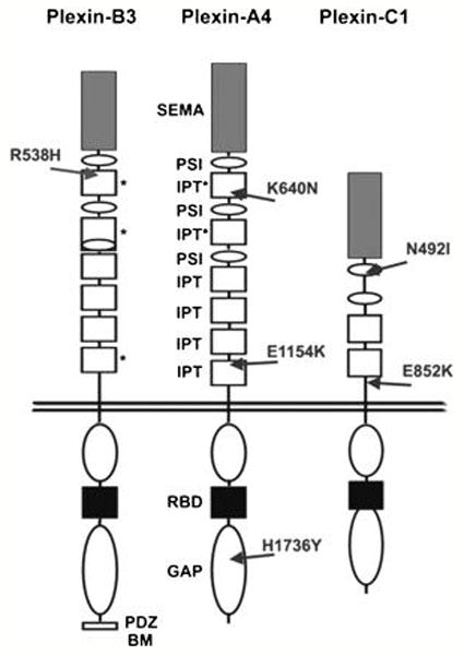
Figure 3. Schematic representation of plexin domain structure and mutations.

The location of novel missense mutations found in human cancer samples are indicated by arrows. In the extracellular portions, grey boxes with “SEMA” indicate semaphorin domains, white ovals with “PSI” indicate Plexin-Semaphorin-Integrin domains (also known as MRS motifs), and white boxes with “IPT” indicate Integrin-Plexin-Transcription-factor domains. Previously unidentified IPT-like domains described in this work are marked by asterisks. In the cytoplasmic portion of the receptors, white ovals indicate the conserved GAP-like regions, the black boxes indicate the Rac-Rnd GTP-ase Binding Domains (RBD), while the C-terminus of PlexinB3 includes a PDZ-domain Binding Motif.
