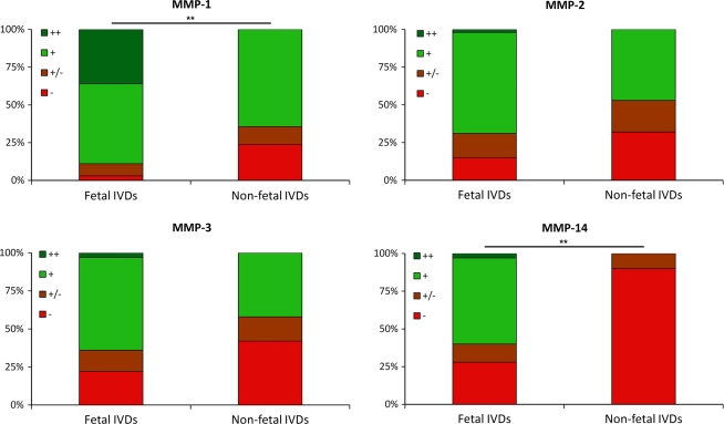
Fig. 3.
Intensity of immunohistochemical staining for MMP-1, -2, -3 and -14 in fetal and non-fetal IVDs. An intense staining for the antigen in >50% of the cells was scored as ++, an intense staining in 10–50% of the cells was scored +, ± was scored if the staining was seen in less than 10% of the cells and − was scored when no staining was observed, **p < 0.005

