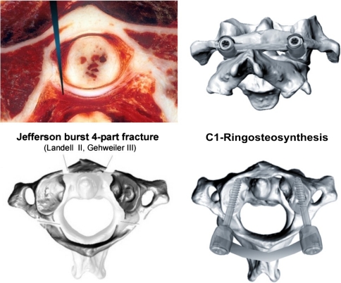
Fig. 1.
Top image on the left cryosection at C1 revealing the atlantodental joint and transverse atlantal ligament (TAL). The long arrow head denotes the level of osteotomy performed in our specimens to transect the TAL before the cyclic loading started. The bottom image on the left illustrates the recreation of the Jefferson burst fracture (JBF) model with the fracture traces being characteristic for JBF. In the current study, a classic four-part JBF was created classified as type II according to Landell [38] and type III according to Gehweiler [22]. The images on the right reveal the C1-ring osteosynthesis with 3.5-mm shaft-screws placed into the C1-lateral mass and linked with a 3.5-mm rod

