
Fig. 1.
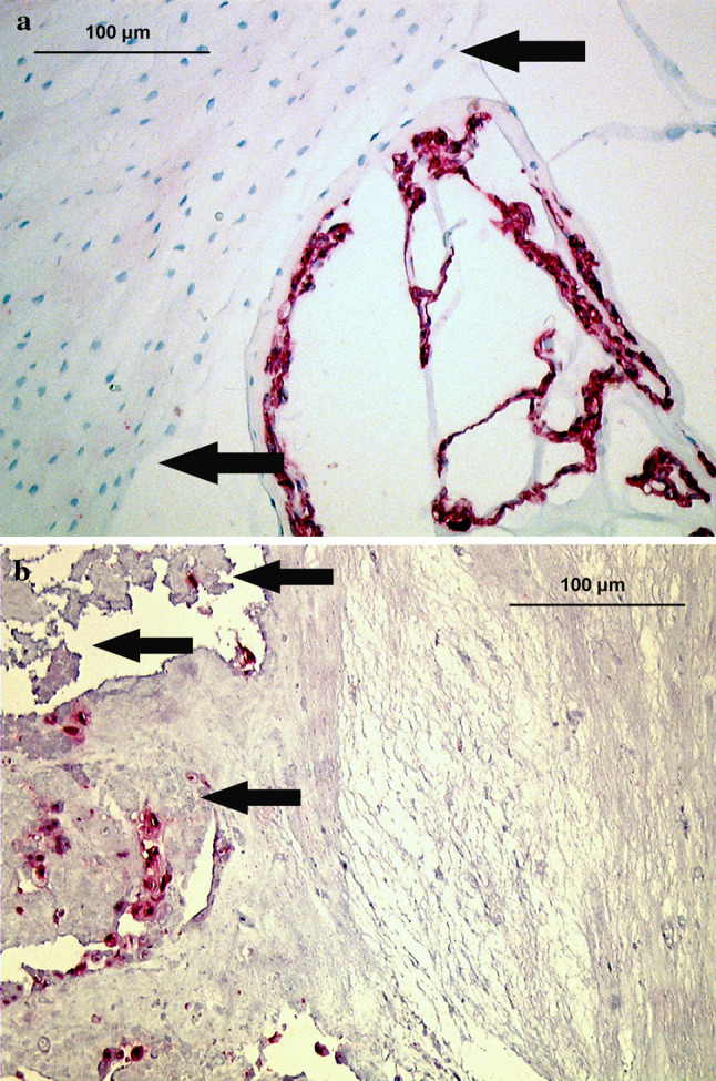
Immunolocalization of pan CK AE1/AE3 (staining cytokeratin-8 and -19 among others) in disc tissue of various age groups (autopsy group). a Fetal nucleus pulposus and pre-anulus region (arrows), showing typical labeling of notochordal cells (28th week, disc level L3/4). b Young adult nucleus pulposus tissue (21 years, disc level L2/3), showing numerous positively stained cells that are exclusively labeled in granular matrix areas (arrows) and adjacent to matrix defects (×200)