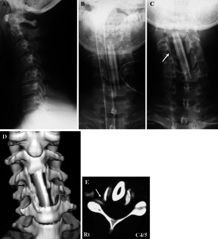
Fig. 2.
Case 2. A preoperative lateral cervical radiograph showing kyphotic alignment of the cervical spine (a). Anterior–posterior views of cervical radiographs just after anterior corpectomy of C4 and C5 and arthrodesis at C3–6 (b) and on the seventh day after surgery (c). c The lateral tilting angle of the grafted fibula was 14° and the right C4–C5 uncovertebral joint was subluxed (arrow). Front view of three-dimensional CT (d) and axial CT images at the level of C4–C5 (e). CT 8 weeks postoperatively showing a subluxed right C4–C5 uncovertebral joint and stenosis of the right C4–C5 foramen (arrows)

