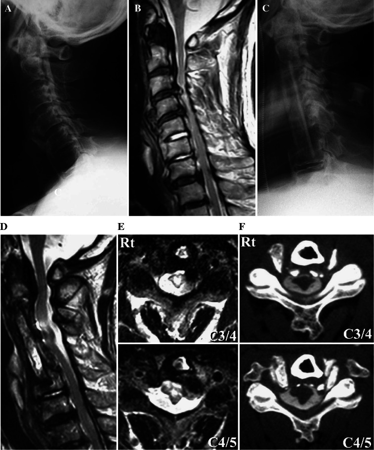
Fig. 3.
Case 9. A preoperative lateral cervical radiograph (a) showing mixed type OPLL from C1 to C6. A midsagittal T2-weighted MR image (b) showing severe compression of the spinal cord and HSCs at C3–C4 and C4–C5 levels. A postoperative lateral cervical radiograph shows anterior corpectomy of C3, C4, and C5 and arthrodesis at C2–C6 (c). T2-weighted MR midsagittal (d) and axial views at C3–C4 and C4–C5 (e) and a CT myelogram (f) showing an excessive anterior shift of the spinal cord at C3–C5. e HSCs in the gray matter at the C3–C4 and C4–C5 levels

