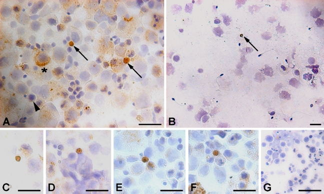
Figure 4.
Immunocytochemical localization of Oct-4A and Oct-4B in testicular germ cell smears. A distinct cell population stained positive using polyclonal antibody (Abcam) with nuclear Oct-4A and a characteristic unstained rim of cytoplasm (arrow, A). These cells were few in number, thus different fields were captured to demonstrate their presence (C–F). Another population comprised large-sized cells with cytoplasmic Oct-4B (asterisk, A). A distinct gradation of staining intensity for Oct-4B was observed in the cytoplasm of these cells, ranging from dark brown to no stain (arrowhead). Only nuclear Oct-4-positive cells (arrow) were detected using MAb from Santa Cruz (B), and no stain was observed in negative control (G). Bar = 20 μm.

