Figure 1.
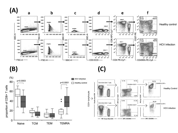
Distribution of CD8+ T cell subsets in HCV-infected patients and healthy controls according to the expression of CD45RA and CD27. (A). Representative Dot plots analysis showing the gating strategy to define CD8+ T cell subsets using CD45RA and CD27. The plots from (a) to (f) were first gated on lymphocytes by FSC and SSC, then the CD3+CD4-CD8+ subpopulation was defined by the expression of CD45RA and CD27. (B). Comparison of CD8+ T cell subsets in chronic HCV infection (dark grey boxes) and healthy controls (open boxes). Data were shown as median and interquartile range values. Symbols: ●, outlier values (more than 1.5 times the interquartile range). (C). Dot plots analysis showing that the majority of CD8+ CD45RA+CD27+ naïve T cells presented CD45RA+CCR7+ phenotype. A representative dot plot result of five HCV patients and five healthy donors,

