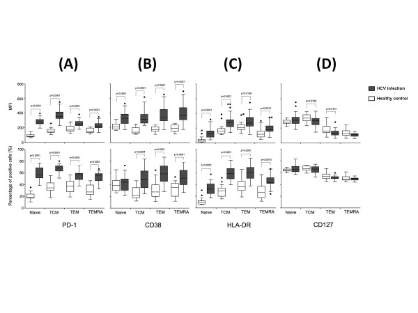
Figure 3.
Comparison of PD-1(A), CD38 (B), HLA-DR(C) and CD127 (D) expression on CD8+ T cell subsets (naïve, TCM, TEM and TEMRA). Data were presented as MFI (top panel) and percentage of positive cells (low panel) between chronic HCV infection (dark grey boxes) and healthy controls (open boxes). Graphs showed median and interquartile range values of MFI or of percentages of positive cells. Symbols:●, outlier values (more than1.5 times the interquartile range).

