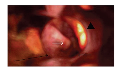
Figure 1.
EUA. Direct hypopharyngoscopy, white arrow shows the bulge of the posterior hypopharyngeal wall into left piriform sinus. The long black arrow shows the posterior pharyngeal wall while the head of an arrow demonstrates the epiglottis. The short black arrow shows small part of the glottis marginally visible through the direct endoscope.

