
Figure 1.
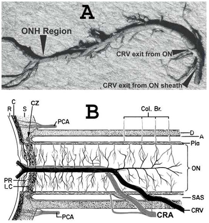
(A) Cast of the central retinal vein showing its entire course from the optic disc to its exit from the optic nerve sheath. Note the presence of a large number of prominent collaterals within the optic nerve and none in the optic nerve head region in this specimen.
(B). Schematic representation of the blood vessels in the optic nerve. (B modified from Hayreh SS: Trans Am Acad Ophthalmol Otolaryngol 1974; 78:OP240-OP254.)
Abbreviations: A = arachnoid; C = choroid; CAR and CRA = central retinal artery; Col. Br. = Collateral branches; CRV = central retinal vein; CZ = circle of Zinn and Haller; D = dura; LC = lamina cribrosa; ON = optic nerve; ONH = optic nerve head; PCA = posterior ciliary artery; PR = prelaminar region; R = retina; Rec. Br. CZ = recurrent pial branches from peripapillary choroid/CZ; S = sclera; SAS = subarachnoid space.