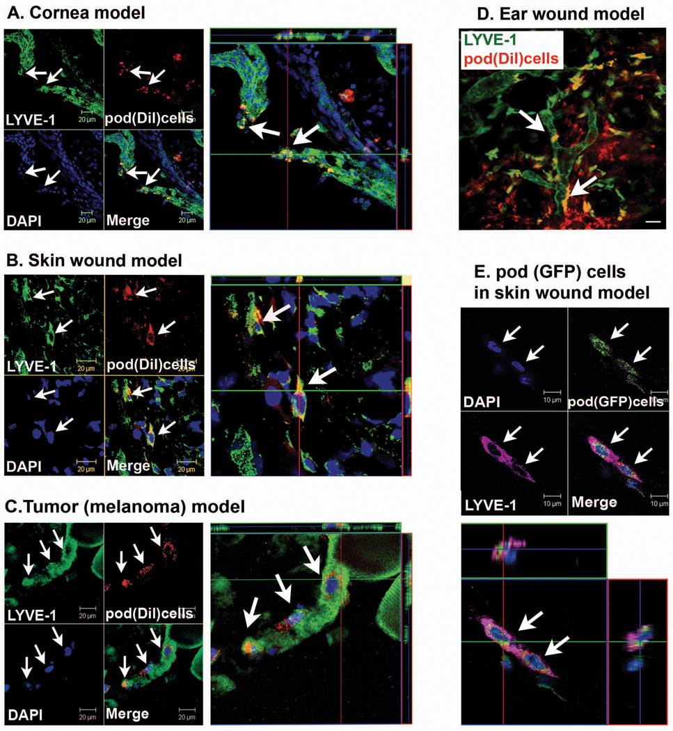
Figure 3. Lymphvasculogenesis from pod+ cells in animal models.
A through C, Mice which had received surgery for cornea micropocket, wound or implantation of tumor cells (B16-F1 Melanoma) were injected with Dil-labeled pod+ cells (red) and the tissues were harvested 7 days later for immunohistochemistry. A through C, Representative confocal images from cornea (A), wounded skin (B) and peritumoral subcutaneous tissues (C) demonstrated that DiI-labeled pod+ cells were incorporated into lymphatic vessels and exhibited a LEC marker, LYVE-1. D, In vivo live confocal microscopic image from an ear wound model showed that multiple pod+ cells (DiI) were clearly incorporated into lymphatic vessels and colocalized with LYVE-1. Arrows indicate cells positive for Dil and LYVE-1. Scale bar = 20 µm. E, Skin wound tissues injected with pod+ cells from GFP mice were stained for LYVE-1 and examined by confocal microscopy. Injected pod+ GFP cells (arrows) were incorporated into the lymphatic vessels and expressed LYVE-1. Representative images from at least two independent experiments for each animal model are shown (n = 3 per experiment). Scale bar = 10 µm. Blue fluorescence: DAPI

