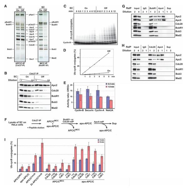
Fig. 1.
Biochemical characterization of APC/CMCC and apo-APC/C. (A) SDS-PAGE–silver-staining analysis of APC/C and MCC from cells in which the checkpoint was active (SC on) or inhibited (SC off). (B) Semiquantitative immunoblot analysis of APC/C from cells in which the checkpoint was on or off. (C and D) Cyclin B ubiquitylation activity of APC/C isolated from cells in which the checkpoint was on or off, analyzed with SDS-PAGE and autoradiography (C) and quantified (D). (E)Checkpoint activation reduces APC/C activity similarly toward prometaphase (cyclin A and Nek2A) and metaphase substrates (cyclin B and securin). Ratios of ubiquitiylation by the two APC/C samples were calculated for each substrate in three independent experiments. Error bars indicate SD of the mean. (F) Schematic representation of APC/CMCC and apo-APC/C isolation from cells with an active checkpoint (reIP, reimmunoprecipitation). (G and H) Immunoblot analyses of complexes obtained in (F). (I) Cyclin B ubiquitylation activities of APC/CCdc20 from cells with an inactive checkpoint and of APC/CMCC and apo-APC/C from cells with an active checkpoint. APC/C levels were normalized to Apc2. APC/CMCC and apo-APC/C were preincubated with various concentrations of recombinant Cdc20 or Cdh1. Cyclin B ubiquitylation was quantified after 4- and 8-min reaction times. Data represent three independent experiments and error bars indicate SD of the mean.

