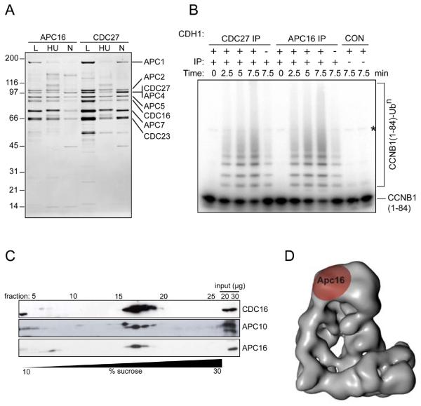
Fig. 4.
Characterization of APC16, a previously unknown subunit of the APC/C.
(A) Silver-stained SDS-PAGE gel showing proteins immunoprecipitated using APC16 or CDC27 antibodies from extracts of HeLa cells, cultured under logarithmic growth conditions (L), or arrested in S phase by treatment for 18 hr with hydroxyurea (HU) or arrested in prometaphase by treatment for 18 hr with nocodazole (N). Numbers on the left indicate the molecular masses of reference proteins.
(B) Phosphorimage showing the ubiquitylation of Cyclin B1 (CCNB1), catalyzed by CDC27 and APC16 immunoprecipitates. [125I]-labelled human CCNB1 fragment (amino acids 1 to 84) was incubated with CDC27 or APC16 immunoprecipitates from logarithmically growing HeLa cells, plus E1 and E2 enzymes, ubiquitin and ATP, with or without the recombinant co-activator protein CDH1, for the times indicated, then analyzed by SDS-PAGE and phosphorimaging. CON indicates empty protein-A beads (left) and a condensin antibody immunoprecipitate (right). The asterisk marks a contaminating band present in the CCNB1 sample.
(C) Immunoblots showing the co-sedimentation of APC16 with core APC/C subunits CDC16 and APC10, following density gradient centrifugation. An extract of logarithmically-growing HeLa cells was subjected to centrifugation through a 10-30% sucrose density gradient. 28 fractions were collected and analyzed by SDS-PAGE and immunoblotting with the antibodies indicated.
(D) Three-dimensional model of the human APC/C obtained by electron microscopy (30), showing the location of APC16, as determined by antibody labeling.

