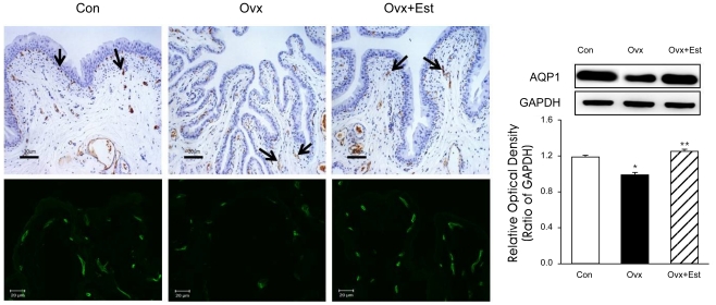
Figure 2.
Immunohistochemistry of AQP1 in urinary bladder tissue from animals of the control (Con), ovariectomy (Ovx), and ovariectomy plus 17β-estradiol treatment (Ovx+Est) groups. Immunolabeling of AQP appears in brown (arrows). AQP1 was mainly expressed in the capillaries and venules. The panels below are displayed at immunofluorescence stain in each group. The anti-AQP antibodies recognized 27-29 kDa bands, corresponding to glycosylated AQPs. Anti-actin antibody recognized the 42 kDa band. AQP-1 protein was present in all groups, and there were no significant changes among the groups. The right panels denote the means±standard deviation of 10 experiments for each condition determined by densitometry relative to GAPDH. *p<0.05 vs. control. **p<0.05 vs. Ovx. AQP=aquaporin; Con=control; Ovx=ovariectomy; Ovx+Est=ovariectomy plus 17β-estradiol treatment. Horizontal scale bar at the bottom of each figure indicates the magnification power.

