
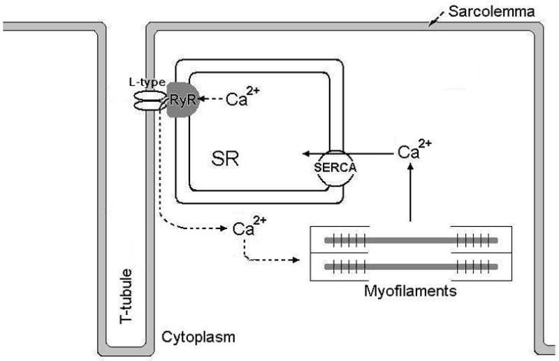
Figure 1. Excitation-contraction coupling in cardiac and skeletal muscle.
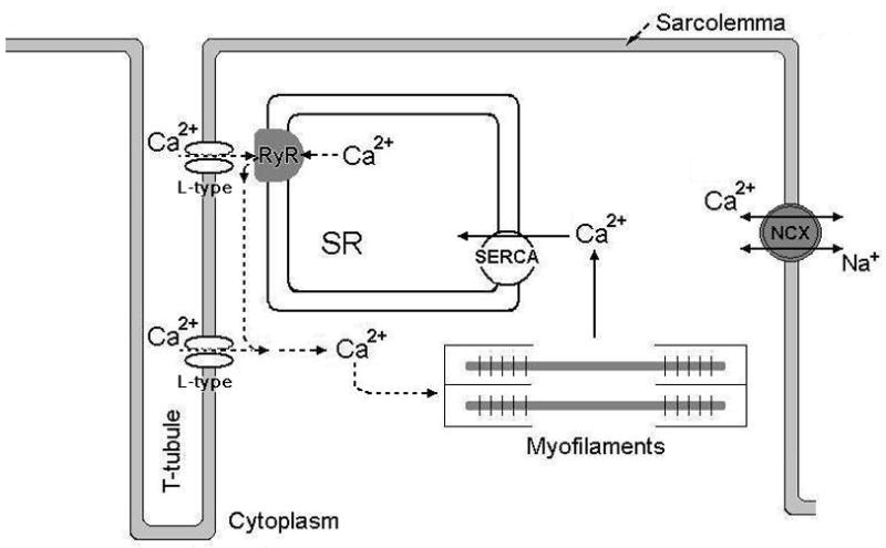
A) Figure showing the principals of Ca2+ cycling in excitation-contraction (E-C) coupling of the cardiomyocyte. Upon depolarization of the sarcolemma, L-type Ca2+ channels open to allow Ca2+ to enter the cell. This Ca2+ will in turn activate the juxtaposed RyR and more Ca2+ will be released. The bulk of cytoplasmic [Ca2+] increase is due to RyR-mediated release of Ca2+ from the SR. Cytoplasmic Ca2+ will trigger myofilament activity so that contraction can occur. To relax, cytoplasmic Ca2+ is pumped back into the SR via the SR Ca2+ ATPase (SERCA2a) and extruded out of the cell through the sarcolemmal Na+-Ca2+ exchanger (NCX).
B) Depicts Ca2+ handling in skeletal muscle E-C coupling. L-type Ca2+ channels at the sarcolemma and RyR1 in the SR membrane are in close vicinity with each other. Depolarization of the sarcolemma causes the L-type Ca2+ channel to interact with and activate the RyR independent of entry of extracellular Ca2+. Virtually all of the increase in cytoplasmic [Ca2+] that is needed for myofilament activation and force generation comes from SR Ca2+ release. The Ca2+ is cleared from the cytoplasm via the SERCA. The absence of transsarcolemmal Ca2+ fluxes underscores the importance of RyR1 Ca2+ release function in skeletal muscle ECC.