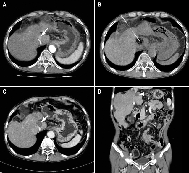
Fig. 2.
Computed tomography (CT)-guided high-dose percutaneous ethanol injection (PEI) therapy in a 53-year-old man with a small hepatocellular carcinoma in the caudate of the liver that abutted on inferior vena cava and was invisible in ultrasound. (A) Contrast-enhanced CT image shows a mass in the caudate lobe of the liver (arrow). (B) CT-guided PEI therapy was performed for this mass at two sessions, with 20 mL of 99% ethanol being injected during each session. (C, D) Contrast-enhanced axial (C) and coronal (D) abdominal CT images obtained 3 years after PEI therapy show complete necrosis of this mass (arrows).

