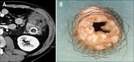
Fig. 11.
A metallic stent had been placed in the descending colon of a 54-year-old patient 6 months earlier. (A) Computed tomography axial image shows a large tumoral mass (asterisk) in the descending colon with occlusion of the stent lumen. (B) Surgical specimen shows the Wallstent with occlusion of the lumen due to tumor ingrowth.

